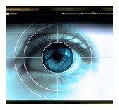
Retina Exam
The function of the eye’s retina can be compared to film in a camera
– it sends light rays and raw sensory information to the brain via the
optic nerve. These electric signals are then translated into vibrant and
colorful images, or what we call “sight.”
The retinal membrane
covers the inside of the back of the eye. In it are light-sensitive
nerve cells called rods, and cones, which discern both light and color.
Damage or disease in the retina can lead to impaired vision and in some
cases, blindness.
Although your primary eye doctor can detect signs of retina
disorders, most people are referred to a retina specialist. These
doctors are specially trained to test for and treat retinal disease or
injury. Early detection is critical because it can be difficult to
repair the retina once it has been damaged.
What to Expect
First your retina specialist will take your medical history and
create your patient record. It is a good idea to bring a list of any
medications you are currently taking. You should also bring a pair of
sunglasses, since you will have your eyes dilated. We also recommend
that you have someone drive you to your appointment, because the pupil
dilation will make it hard for you to focus and affect your ability to
drive.
During the actual exam, your doctor will conduct a few
different tests. The length of the appointment is typically 2-3 hours.
Some of the diagnostic tests performed include the following:
- Indirect Ophthalmoscopy: This gives your doctor a comprehensive
view of the internal structures of your eyes. A small handheld lens
and a light attached to a headband are used to examine the inside of
your eye and get a peripheral view of the retina.
- Visual Field Testing: During this test, you will be asked to
focus on a point straight ahead. Then, flashes of light are
displayed on a screen and you press a button whenever you see one of
the flashes. A computer records the results, constructing a virtual
map of your visual field. Your doctor will look for “blind spots,”
which are a potential sign of retinal disease.
- Ultrasound: A-scan and B-scan ultrasounds are helpful diagnostic
tools that provide a 2-D cross-sectional view of the eye. The test
is quick and painless, using high frequency sound waves to produce a
topographical view of the inner eye structures.
- Fluorescein Angiography: This test allows your doctor to
evaluate the blood vessels in the retina. During the test, a
vegetable-based dye is injected into your arm. Once it reaches the
vessels in the eyes, it will glow in visible light and images will
be recorded. Certain eye disorders cause poor retinal circulation,
which this test can help detect.
- Fundus Photography: Before this procedure, your pupils are
dilated to prevent them from constricting. A special camera mounted
to a microscope is used to capture images of the eye’s internal
structures. While the pictures are being taken, you will see a
series of bright flashes. These images help document any retinal
abnormalities, as well as monitor treatment. The entire process
takes five to ten minutes.
After the Exam
Following your retina exam, your doctor will discuss the results with
you. He or she will determine if further testing is necessary, or if a
diagnosis has been established. After explaining your treatment options,
your doctor will address any questions you may have and make
recommendations for future care.
Early detection and
comprehensive care are vital to your optical health, and we are
dedicated to providing both for our patients. If you live in or around
Boston please do not hesitate to
call us if you have any questions about your retina exam.
Go back to Patient Information
|